
Embark on a journey through the fascinating world of radiography as we unravel the secrets behind image formation. Imagine peering inside the human body, exploring the dance of photons, and deciphering the language of contrast that brings diagnostic images to life.
This isn’t just about X-rays; it’s a captivating exploration of the art and science that transforms beams of light into windows, revealing the hidden wonders within us. Join us as we navigate the complexities of contrast, discover the nuances of clarity, and understand how the principles of radiographic imaging become the pillars of accurate diagnoses in medical diagnostics!
Table of Contents
The Foundation of Radiographic Imaging
Let’s start at the beginning – the foundational principles that govern radiographic imaging. From X-rays to digital technology, grasp the core concepts that lay the groundwork for accurate diagnostic magic.
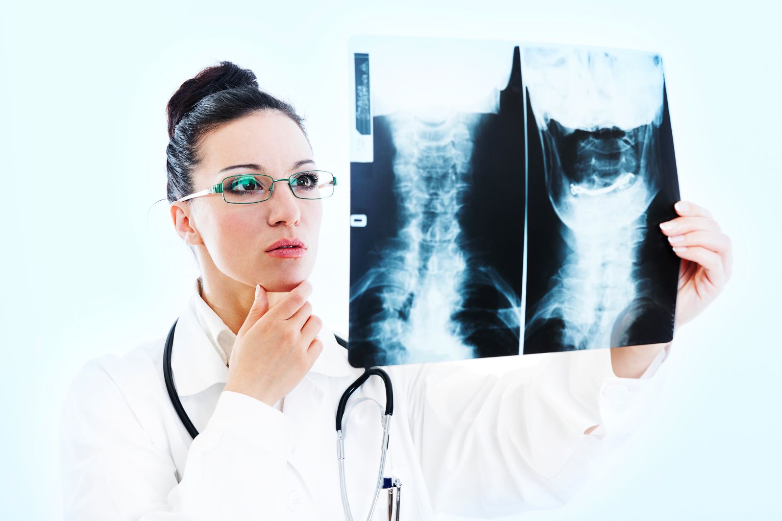
X-ray Generation
To start seeing inside the body, we use something called X-rays. These are like special beams of light that can go through us and take pictures of what’s inside. It’s like a sneak peek to see our bones and organs. The X-rays act like messengers, going through the body and capturing pictures of what’s inside. In this way, X-rays in Lake Hopatcong, NJ and other areas can be used to accurately diagnose issues that might otherwise go undetected.
Attenuation and Contrast
When the X-rays travel through our body, some parts slow them down more than others. We call this slowing down “attenuation.” Because different parts slow down the X-rays differently, the pictures show different shades and contrasts. It’s like having various colors in a picture, making it easier to see details.
Anatomy and Proper Positioning
To get the right pictures, we need to position ourselves correctly. It’s like taking a good photo – if you’re in the right place, the picture will appear better. So, we must ensure the person being scanned is in the proper position and the X-ray machine is set up correctly.
The interplay of X-rays with Tissues
The X-rays interact with our body’s parts, creating detailed pictures. It’s like the X-rays and our body parts are talking to each other. Understanding how different body parts react to X-rays helps us see things clearly in the pictures. It can also be a starting point if you want to continue your education in radiology, taking your understanding to a whole new level.
Different Imaging Techniques
Several different X-ray techniques are used in medical imaging, each tailored to specific diagnostic needs. Here are some standard X-ray techniques:
Plain Radiography (Conventional X-rays)
Plain radiography, or conventional X-rays, is a primary way of taking pictures inside the body. It sends X-rays through the body to make a simple picture of bones and organs. Doctors often use this method first to check for fractures or bone problems. It’s like taking a quick snapshot to get an initial look at what’s happening inside.
Fluoroscopy
Fluoroscopy is like a live X-ray movie that continuously takes pictures in real time. It’s handy for seeing things in motion inside the body, like how organs move or tracking contrast agents as they travel through blood vessels.
Fluoroscopy gives doctors helpful information about how organs and structures are working. It’s like watching a video instead of just looking at a snapshot, providing a dynamic view for better understanding.
Computed Tomography (CT Scan)
A CT scan, short for Computed Tomography, is a unique X-ray technique that takes detailed pictures of the inside of the body in slices. It’s like making a layered cake where each slice shows different parts. This method captures images of soft tissues and organs, helping doctors diagnose various health issues such as tumors or internal injuries.
Mammography
Mammography is a special X-ray used to take pictures of breast tissue. It’s like a dedicated tool for checking how the breasts are doing. The main job of mammography is to help catch signs of breast cancer early.
By taking detailed images, it assists doctors in spotting any abnormalities or potential tumors in the breast. This technique is crucial for early detection, allowing for timely and effective treatment if any concerns are found. In a way, it’s like a proactive step in maintaining breast health.
Dual-energy X-ray Absorptiometry (DEXA or DXA)
Dual-energy X-ray Absorptiometry, or DEXA, is used to check how dense (strong) our bones are. It works by using low-dose X-rays to measure the minerals in our bones. This helps doctors understand the bone’s strength and can be helpful in diagnosing and managing conditions like osteoporosis.
Cone Beam CT (CBCT)
Cone Beam CT is a unique way of taking pictures that uses a cone-shaped X-ray beam to create detailed 3D images. It’s like capturing a complete and thorough view of a specific area. This technique is commonly used in dental and maxillofacial (related to the jaw and face) imaging.
Positron Emission Tomography-Computed Tomography (PET-CT)
PET-CT is a powerful combination of nuclear medicine and CT imaging. It gives us functional and anatomical information about the inside of the body. It’s like having a comprehensive view that shows the structure and how things are working.
This technique is beneficial in finding out about the body’s metabolic activity. This is crucial for diagnosing and understanding the stages of different medical conditions, including cancer.
Digital Radiography
In today’s medical settings, digital radiography is widely used because it makes the process more efficient. Instead of using film, it uses digital detectors to capture images of the inside of the body.
Plus, it helps reduce the amount of radiation patients are exposed to. It’s like bringing the benefits of digital technology to make medical imaging better and safer.
Pneumocolon (Air Contrast Barium Enema)
Pneumocolon, or air contrast barium enema, is a method to see inside the colon using X-rays. During this technique, a combination of barium sulfate and air is used to highlight the details of the colon in the X-ray images. By doing this, doctors can better diagnose conditions affecting the colon.
Unveiling the Principles of Radiographic Imaging
They apply the fundamental principles of radiographic imaging, namely proper posis crucial for achieving accurate diagnoses. Healthcare professionals must continue prioritizing these aspects to deliver high-quality patient care.
Elevate your diagnostic skills by embracing the magic of radiographic imaging principles. Let us strive to consistently adhere to these principles and improve patient outcomes. Don’t wait- start implementing them in your practice today!
Ready for more insights? Explore the rest of our site today!
